Figure 1. (Click to enlarge) |
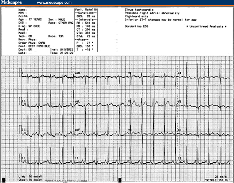 Figure 2. (Click to enlarge) |
 Figure 3. (Click to enlarge) |
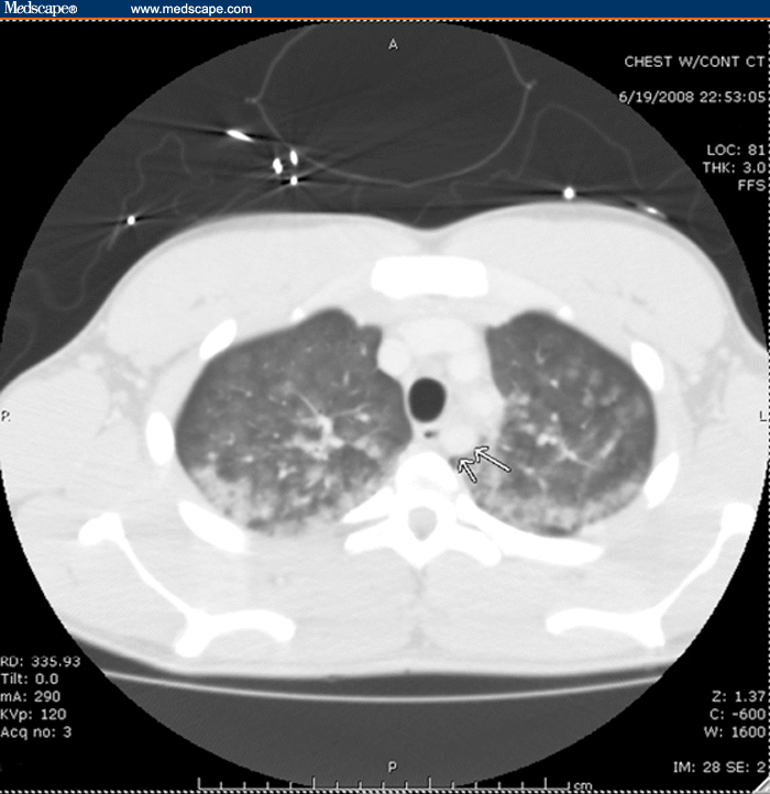 Figure 4. (Click to enlarge) |
A paramedic call is received in the emergency department (ED) reporting a 10-min estimated time of arrival for a 17-year-old male who was found in cardiac arrest following a blow to the chest. The patient has regained spontaneous circulation, and is currently stable and maintaining his own airway. A rhythm strip faxed to the ED prior to the patient's arrival shows ventricular fibrillation, with subsequent conversion to a normal sinus rhythm after defibrillation with 200 joules (see Figure 1). Further questioning of the prehospital personnel reveals a history of witnesses reporting that the patient, a center outfielder for a local baseball team, was trying to catch a baseball when one of his teammates accidentally ran into him, elbowing him in the middle of his chest. The patient immediately dropped to the ground and was unresponsive. Cardiopulmonary resuscitation (CPR) was initiated by his coach after no pulses were palpated. The paramedics arrived 5 minutes later and, as noted on the rhythm strip, found the patient to be in ventricular fibrillation. One 200 joule countershock was administered, converting the ventricular fibrillation to a normal sinus rhythm, and the patient was noted to regain consciousness.
Upon arrival at the ED, the patient reports only mild anterior chest wall pain and denies any substernal chest pain, shortness of breath, palpitations, weakness, or confusion. He states that he has never before fainted. The patient and his mother deny any significant past medical or family history, including any arrhythmias, unexplained sudden deaths, or cardiac structural diseases. He denies having a lower exercise tolerance than his teammates and also denies any smoking, drinking, use of medications, illicit substance abuse, or doping practices.
On physical examination, the primary survey of his airway, breathing, and circulation is unremarkable. The patient has a blood pressure of 130/71 mm Hg and a heart rate of 106 bpm, with a normal rhythm. The respirations are 28-30 breaths/min. The initial oxygen saturation is 83% while the patient is breathing room air, but it corrects to 98% on a non-rebreather mask and, subsequently, to 99% on 2 L nasal cannula. His mentation is intact and he remains alert, with a Glasgow Coma Scale rating of 15. The skin examination reveals mild ecchymosis just anterior to his sternum. The lungs are clear to auscultation bilaterally, and the cardiac examination reveals a regular rate, with normal S1 and S2 heart sounds and no clicks, gallops, rubs, or murmurs. The abdominal and neurologic examinations are unremarkable. The musculoskeletal exam shows the patient's wingspan to be 95-100% of his height.
The patient is placed on a cardiac monitor upon arrival to the ED. A 12-lead electrocardiogram (ECG) is obtained that reveals sinus tachycardia at a rate of 110 bpm, with mild right-axis deviation. The QRS complex, QT interval, ST/T waves, and P waves are all noted to be normal (see Figure 2). A portable, upright chest radiograph shows somewhat underaerated lungs but no signs of fractures, widening of the mediastinum, cardiomegaly, or hemopneumothorax (see Figure 3). A complete blood count (CBC) is normal, except for a mildly elevated white blood cell (WBC) count of 13.6 ×10³/µL (13.6 ×10⁹/L). A metabolic panel is normal, including normal potassium and magnesium findings. The initial troponin I is 0.04 ng/mL (0.04 µg/L; normal range, 0.02-0.04 ng/mL; indeterminate 0.05-0.30 ng/mL). A urine drug screen is negative. Computed tomography (CT) scanning of the chest (see Figure 4) is remarkable only for mild pulmonary and periportal edema. The patient is admitted to the pediatric intensive care unit (PICU) for continuous cardiac monitoring and cardiology consultation. An echocardiogram is ordered in the ED, to be done in the PICU.
What happened here? What is the most likely underlying reason for this young man's cardiac arrest? Why is it important to make the diagnosis and institute the correct form of treatment?
The diagnosis is Commotio Cordis.
Commotio cordis (which is Latin for "disturbance of the heart") is, in essence, a concussion of the heart. Initially described as early as 1857, it is defined as an instantaneous cardiac arrest produced by a witnessed, nonpenetrating blow to the chest, in the absence of preexisting heart disease or identifiable morphologic injury to the sternum, ribs, chest wall, or heart. Commotio cordis is a diagnosis of exclusion in that other causes, such as substance abuse, myocardial infarction, electrolyte abnormality, prolonged QT syndrome, and hypertrophic obstructive cardiomyopathy (HOCM), must first be ruled out with examinations such as urine drug screens, serial assessment of cardiac biomarkers and EKGs, electrolyte level testing, and echocardiography.[1,2]
Although reported as the second most common cause of sudden cardiac arrest in young athletes (behind HOCM), commotio cordis is underreported and underrecognized.[2] The United States Commotio Cordis Registry (USCCR), in Minneapolis, Minnesota, reported that as of September 2001, only 180 cases had been documented. Up to 62% of these cases involved engagement in organized, competitive sports, with two-thirds of the patients being younger than 16 years of age and 80% being male. The oldest reported case was that of a 20-year-old man struck in the chest by a baseball, and the youngest case was that of a 7-week-old crying infant struck in the chest by his frustrated father. Eighty-one percent of cases involved a blunt, precordial blow from a projectile object propelled against a stationary chest wall, resulting in a relatively localized area of contact. It is notable that those who are most susceptible to commotio cordis are young athletic males. This is probably the result of the fact that there is less protection of the heart by subcutaneous fat, muscle bulk, and fully ossified ribs, all of which become more common in adulthood.[2,3] A review of the USCCR data revealed that a vast majority of the 180 reported cases were caused by a blow to the chest from an object used during an organized youth sporting event. A baseball accounted for 53 of the cases, with a softball and a hockey puck following, at 14 and 10 cases, respectively. Other documented sporting cases have been caused by blows delivered by body parts, such as an elbow, knee, foot, or fist hitting the anterior chest wall (5-6 cases of each). Finally, daily activities, including parent-child discipline (5 cases), and even a fall from monkey bars (1 case), can lead to commotio cordis. Regardless of the mechanism, impacts resulting in commotio cordis are typically of low energy and velocity.[1,4] The victim may collapse immediately after the blow, but in up to 50% of cases, there is a short delay between the times of impact and collapse.
In 1930, George Schlomka was the first to describe the factors that can lead to arrhythmia after a moderate precordial impact. He believed that the force, location, and type of object causing the impact determined the type of injury and the subsequent risk of arrhythmia.[2] The force transmitted to the heart is directly related to the hardness of the striking object. Madia et al reported that the threshold speed of impact at which a standard baseball can cause ventricular fibrillation is between 25 and 30mph. When the speed is over 50mph, however, the likelihood of ventricular fibrillation actually decreases, although the possibility of myocardial contusion becomes greater. Furthermore, the authors stated that the impact must be directly over the cardiac silhouette near or just to the left of the sternum in order to instigate ventricular fibrillation. Impact on the center of the cardiac silhouette induced ventricular fibrillation in 30% of reported cases, compared with 13% and 4% at the left ventricular base and apex, respectively.[4]
The utilization of a standard baseball leads to the incidence rate as above, but if the core of the ball is softer, then the rate for ventricular fibrillation drops. A study published by Link et al reported that changes to the cores of baseballs to make them softer led to a decrease in the rate of ventricular fibrillation with commotio cordis from 70% to 19%. As such, the use of safety baseballs with rubber cores of different degrees of hardness has been advocated in order to reduce the risk of such traumatic injury in young athletes.[3,5]
Not all impacts to the anterior chest will lead to the ventricular fibrillation observed in commotio cordis. The impact must be delivered 10-30 milliseconds before the peak of the T wave in the cardiac cycle (see Figure 5) in order to induce ventricular fibrillation. Induction is likely secondary to the activation of potassium-carrying ion channels via mechanoelectric coupling. The activation of these ion channels generates an inward current, thus locally augmenting repolarization and resulting in premature ventricular depolarization and the initiation of unstable ventricular arrhythmias. If impact occurs during other portions of the cardiac cycle, different conduction disturbances, such as heart block, bundle branch block, or transient ST segment elevation, may be induced.[2,4,5]
Regardless of etiology, if a young athlete goes into sudden cardiac arrest, CPR should be implemented immediately. Of sports-related cases of commotio cordis documented in the USCCR, 15% of patients survived. In cases in which CPR was instituted within 3 minutes of the impact, 68% of patients survived; however, if CPR was delayed by more than 3 minutes, only 3% of patients survived. Animal studies have shown that CPR instituted within the first 3 minutes of injury can increase survival rate by up to 25%. Concomitantly, early utilization of an automatic external defibrillator (AED) device has been proven to increase survival rates. With an AED recognizing ventricular fibrillation at a sensitivity of 98% and a specificity of 100%, defibrillation within the first 3 minutes can increase the survival rate by an additional 50% or more in animal models, yielding a survival rate of 46% at 4 minutes, and 25% at 6 minutes. The USCCR has recommended that all athletic venues should have an accessible AED. Preventative measures for commotio cordis include parental education, softer baseballs, and protective padding of an athlete's precordium.[1,4] Secondary prevention may involve avoidance of certain sports until the age of 18 years or older.
While the patient in this case was in the PICU, he was placed on continuous cardiac monitoring for 24 hours, and no incident of arrhythmia was noted. An echocardiogram was obtained that revealed a normal left ventricular systolic ejection fraction, with no structural abnormalities, valvular disease, or hypertrophy. A repeat 12-lead ECG showed no changes from the previous one, and subsequently, the serial troponins were measured at 0.25 and 0.04 ng/mL. A pediatric cardiologist consultation was unrevealing. The patient was transferred to the pediatric floor the next day, and he was discharged 2 days after initial admission, with no signs of post-arrest sequelae. The patient's final diagnosis, in conjunction with the ED's suspected admitting diagnosis, was commotio cordis.
-Article Excerpted From eMedicine.com