Fainting in a 24 year old male
- Details
- Published: Monday, 01 December 2008 15:42
Background
|
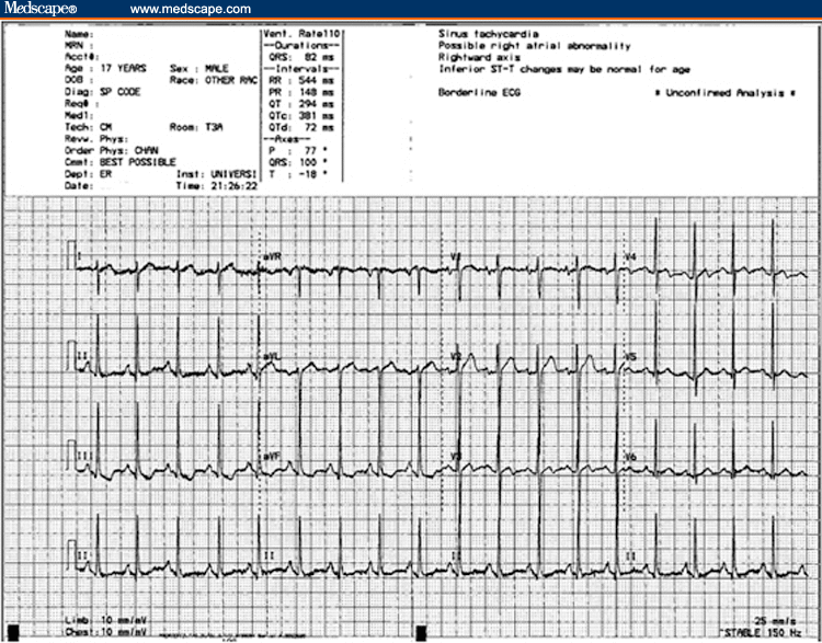
Figure 1. |
A 24-year-old man with no significant past medical history presents to the emergency department (ED) with a complaint of several episodes of a sensation of nearly blacking out. The episodes have occurred about 3-4 times over the 3 days before presentation. The duration of each episode has ranged from a few minutes to over an hour. The patient notes that he has felt his "heart beating really fast," with associated light-headedness. He denies having any chest pain, shortness of breath, or nausea associated with these events. He cannot identify exacerbating or alleviating factors; specifically, he denies exertion as an inciting factor. The remainder of his review of systems is negative except for some mild chronic shortness of breath. The patient takes no medications at home and has no active medical conditions. He smokes 2-4 packs of cigarettes per day and has done so for 5-6 years. He denies any illicit drug use or recent use of over-the-counter medications or herbal remedies. He has no history of any significant cardiac disease or sudden cardiac death in his family.
On physical examination, the patient is afebrile, with a pulse of 65 bpm, a blood pressure of 120/84 mm Hg, and a respiratory rate of 15 breaths/min. His room air saturation reading is 100%. In general, he is well-appearing and in no acute distress. The patient's neck examination shows no jugular venous distention. The heart sounds, including S1and S2, reveal no audible murmurs, rubs, or gallops. The apical impulse is nondisplaced and of normal impact. The lung sounds are diminished throughout, but there are no wheezes, rales, or rhonchi. He has no edema of the lower extremities, and the distal pulses are easily palpable. All other exam findings, including a neurologic examination, are unremarkable.
The patient is placed on a cardiac monitor, and an 18-gauge intravenous (IV) catheter is inserted into the antecubital fossa. Laboratory tests consisting of a complete blood count (CBC) and serum electrolytes are ordered. A portable chest radiograph reveals slight hyperinflation and hyperlucency of the lung fields, with a flattened diaphragm and central pulmonary artery enlargement. An electrocardiogram (ECG) is obtained (see Figure 1).


